7 y.o. FS Portuguese Water Dog with Chronic Vomiting and Diarrhea
History:
-Chronic vomiting, diarrhea, decreased appetite
-Diagnosed with IBD and PLE based on previous endoscopy 3 months prior
-Moderate response to therapy initially, however recurrence of weight loss, lethargy and poor appetite noted frequently.
-Medications: prednisone, metronidazole, famotidine, ondansetron, cyclosporine, mirtazapine
Diagnostics:
CBC: Normal
Chemistry Panel: Elevated liver enzymes, hypoproteinemia and hypoalbuminemia
Previous Diagnostics:
-ACTH Stim: Normal
-Ultrasound: slightly rounded liver with mottling – suspicious for early fibrosis
thickened gastric wall (1cm)
fluid-filled bowel loops and enlarged mesenteric lymph node (3x1cm), ultrasound-guided liver biopsy
-Gastroduodenoscopy:
Stomach: very cobblestoned, edematous appearance. Multiple areas of splotchy red with some erosions – incisura, cardia, fundus all affected.
Duodenum: severely friable and granular. Multiple nodular areas with erosions throughout
-Biopsies
Stomach: Moderate to severe lymphocytic, plasmacytic and neutrophilic gastritis with areas of erosion
Duodenum: Severe, erosive, plasmacytic, lymphocytic, neutrophilic and eosinophilic enteritis with glandular hyperplasia
Moderate cholangiohepatitis with mild multifocal hepatocellular vacuolar change
Problem list:
-History of previously diagnosed IBD and PLE with recurrence of clinical signs
ALICAM Results
-Study Images: 28,797
-Study Time: 13.8h
-Esophageal time: 9 sec (normal)
-Gastric transit time: 6.1h (prolonged)
-SI transit time: 2.1h (normal)
Findings:
-Esophagus: Normal
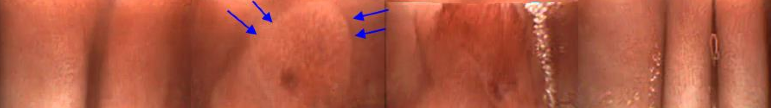
-Stomach: Prolonged gastric transit time. Many areas with erosions, some of which are large, as well as several hematomas and a few nodular areas. In other areas, the mucosa is irregular, with a cobblestone appearance. At the pyloroduodenal junction, linear areas of erosion/ulceration and bleeding are seen.
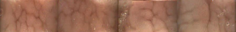
-Small intestine: Diffusely irregular duodenal mucosa with a thickened appearance. On some frames, patches of mucosa look eroded with no villi surrounded by thickened/irregular mucosa that looks nodular. There are rare scattered dilated lacteals seen. The appearance of the mucosa remains irregular throughout the SI, but the severity of the changes lessens as the capsule passes distally.
-Colon: the colonic mucosa is partially obscured by yellow mucoid feces, however, areas with erosion and suspect previous hemorrhage can be seen. The mucosa looks irregular in areas and pale round lesions can be seen in two areas.
Recommendations: View PDF Report

Normal Esophagus
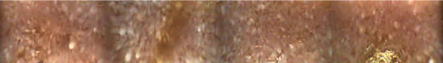
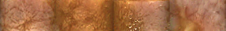
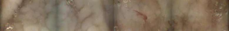
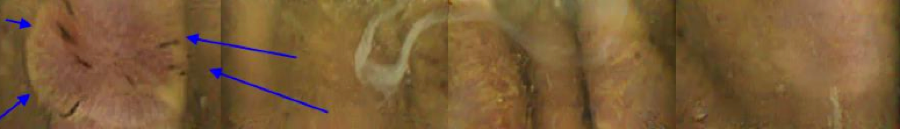
Nodular Area in Gastric Mucosa
Irregular Gastric Mucosa

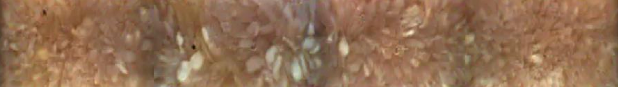
Gastric Erosion Ulcer or Hematoma
Irregular Duodenal Mucosa
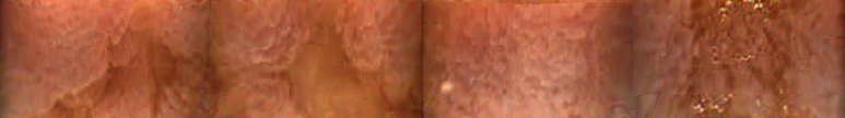
Suspect Eroded SI Mucosa Surrounded by Irregular Thickened Mucosa
Irregular SI Mucosa With a Few Dilated Lacteals
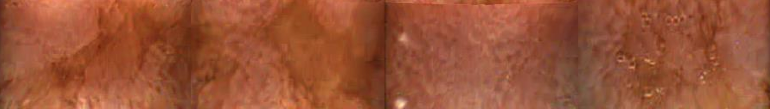
Colonic Erosions


 Gastric erosions with irregular mucosa
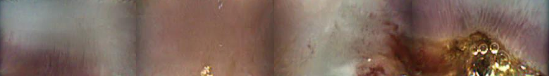 Beginning of Jejunal Mass
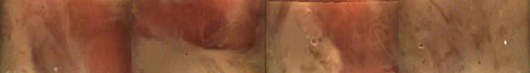
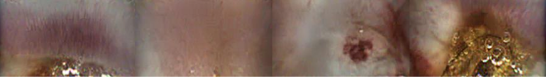 Ulcerated Jejunal Mass
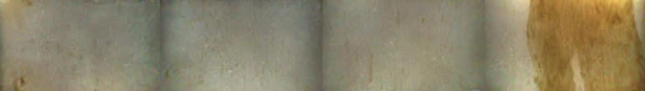
 Gastric Polyp With Bleeding
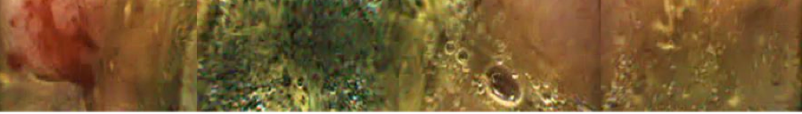 Gastric Polyp With Bleeding 2
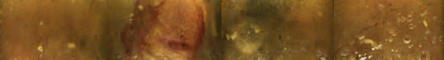 Gastric Polyp With Bleeding 3
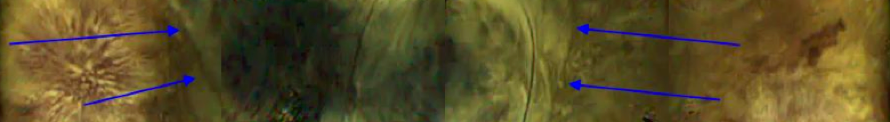 Hematoma
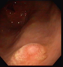
 Gastric Mucosa With Erosion
 Irregular Duodenal Mucosa
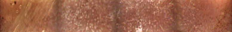 Dilated Lacteals in Duodenum
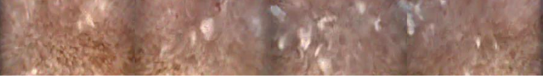 Dilated Lacteals in Jejunum
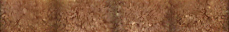 Irregular and Thickened Jejunal Mucosa
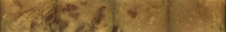 Colon
Colon Irregular and Erythematous Gastric Mucosa
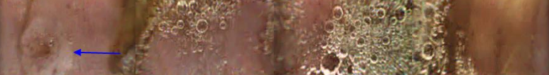 Duodenal Nodular Lesion
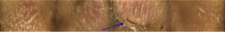
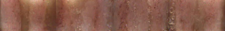 Linear Hemorrhagic Lesions and Irregular Mucosa in Proximal SI
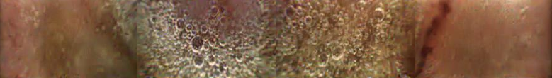 SI Linear Hemorrhagic Lesion
 SI Erosion or Ulcer
 Dilated Lacteals
 Hemorrhagic Lesion in Proximal Colon
 Suspect Pedunculated Gastric Mass
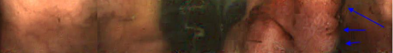 Suspect Gastric Mass
 Irregular Duodenal Mucosa
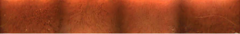 Irregular Thickened Jejunal Mucosa
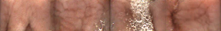 Irregular Gastric Mucosa
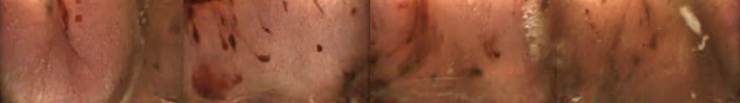 Gastric Erosions
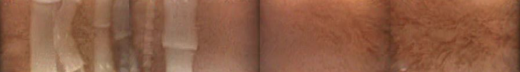 Tapeworms
 Irregular Small Intestinal Mucosa
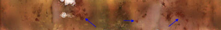 Gastric Erosions Ulcers
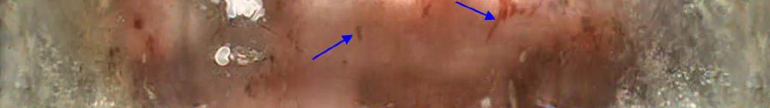 Gastric Erosions